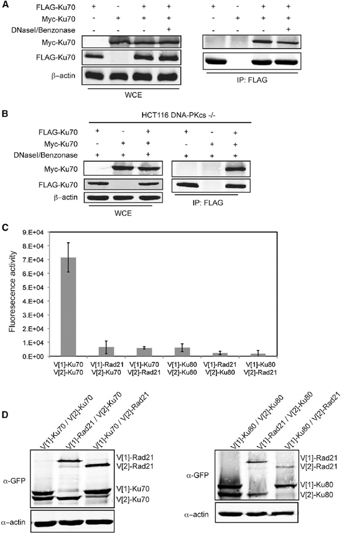
Figure 5. Ku Heterodimers Self-Associate via the Ku70 Subunit.
(A) Coimmunoprecipitation of Myc-Ku70 with FLAG-Ku70. Immunoprecipitations with FLAG antibody were performed using WCEs untreated (−) or treated (+) with DNaseI/Benzonase. FLAG and Myc immunoblots were performed on the WCEs (left) and immunoprecipitates (IP) (right, IP:FLAG). β-actin represents a loading control.
(B) Coimmunoprecipitation of Myc-Ku70 with FLAG-Ku70 in DNaseI/Benzonase treated WCEs from HCT116 DNA-PKcs−/− cells.
(C) Fluorescence quantification of PCA using the indicated combinations of Ku70, Ku80, or Rad21. The error bars represent SD of the mean.
(D) Protein levels resulting from the transfections in (C) detected by immunoblot analysis of WCEs using a GFP antibody. β-actin was used as a loading control.
See also Figure S3.

