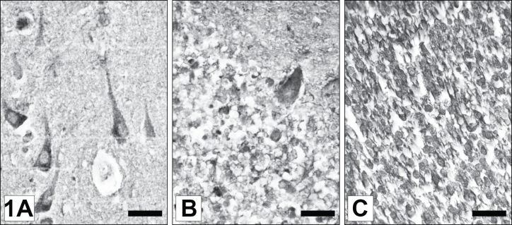
Fig. 1.
Immunohistochemical demonstration of ZNF 804A in adult and developing human brain. (A) Temporal cortex of a 49 years old male. TNF 804A is expressed in layer III pyramidal cells and interneurons. (B) Cerebellum of a 55 years old female. A vast majority of granule cells and a few Purkinje cells were found to express the protein. (C) ZNF 804A expression in the cortex anlage of a fetal brain (19th gestational week). Multiple neuroblasts and radial glial cells are intensely immunostained. Scale Bars (valid for figures 1A–C) = 25 µm.

