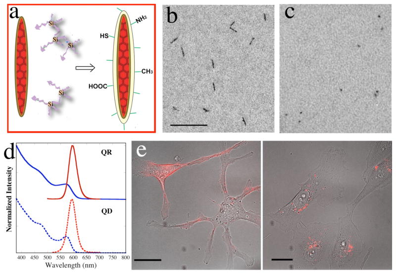
Figure 1.
(a) A cartoon illustrating silanization of quantum rods. Crosslinked silanes are priming molecules for the surface coating. (b) TEM image of silanized rods in neutral phosphate buffer. Scale bar = 100 nm. (c) TEM image of silanized dots in neutral phosphate buffer. Scale bar = 100 nm. (d) The UV-Vis absorption and emission spectra of silanized QR and QD. The blue curves are the absorption spectra; the red curves are the emission spectra. (e) Silanized QRs are biocompatible and non-toxic to living cells. The red fluorescence in the images is from QRs in human breasts cancer cells MDA-MB-231 after 1h (left) and 24h (right) transfected with Chariot™. These are merged images of transmission and fluorescent micrograms. Scale bar is 20 μm.

