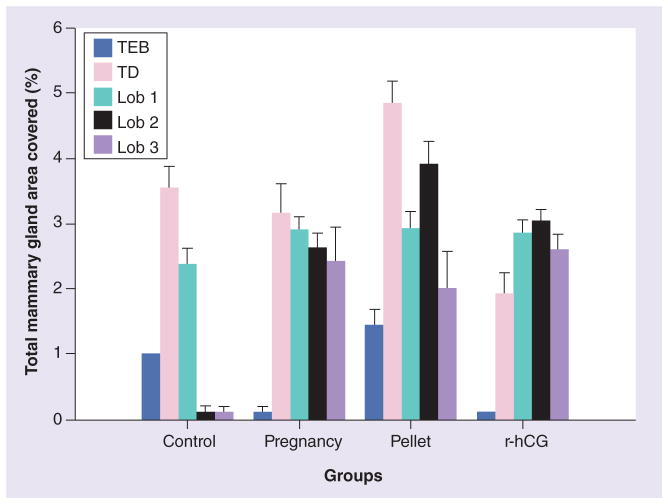
Figure 2. Percentage of structures in the total mammary gland area (mm2) in whole-mount preparations.
The structures on the mammary glands were categorized as TEBs, TDs and lobs 1, 2 and 3. The control rat mammary glands were devoid of lobs 2 and 3, while pregnancy, pellet and r-hCG stimulated the gland differentiation to reach lob 3. None of the rat mammary glands postpregnancy or post-hCG treatment contained TEBs, and they presented lobs 2 and 3, whereas r-hCG presented slightly higher numbers of lobs 2 and 3 compared with pregnancy. The animals treated with the pellet contained – in addition to TDs and lobs 1, 2 and 3 – a greater percentage of TEBs, compared with the control group, indicating that the mammary gland has not been completely differentiated by this treatment. Lob: Lobule type; r-hCG: Recombinant human chorionic gonadotropin; TD: Terminal duct; TEB: Terminal end bud.
Reproduced with permission from Springer Science+Business Media BV [36].

