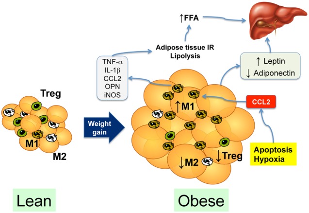
Figure 3. Contribution of the adipose tissue to the development of nonalcoholic steatohepatitis.

In the lean subject, M2-polarized macrophages and regulatory T cells (Treg) predominate in the vascular stromal fraction. Weight gain leads to expansion of the adipose tissue, resulting in tissue hypoxia and adipocyte apoptosis, leading to secretion of chemotactic cytokines such as CCL2 (MCP-1). CCL2 secretion recruits M1-polarized macrophages that secrete proinflammatory cytokines, contributing to insulin resistance, which increases lipolysis and the delivery of free fatty acids (FFA) to the liver. The expanded and inflamed adipose tissue also causes an imbalance in adipokine secretion, with an increase in leptin and a decrease in adiponectin circulating levels.
