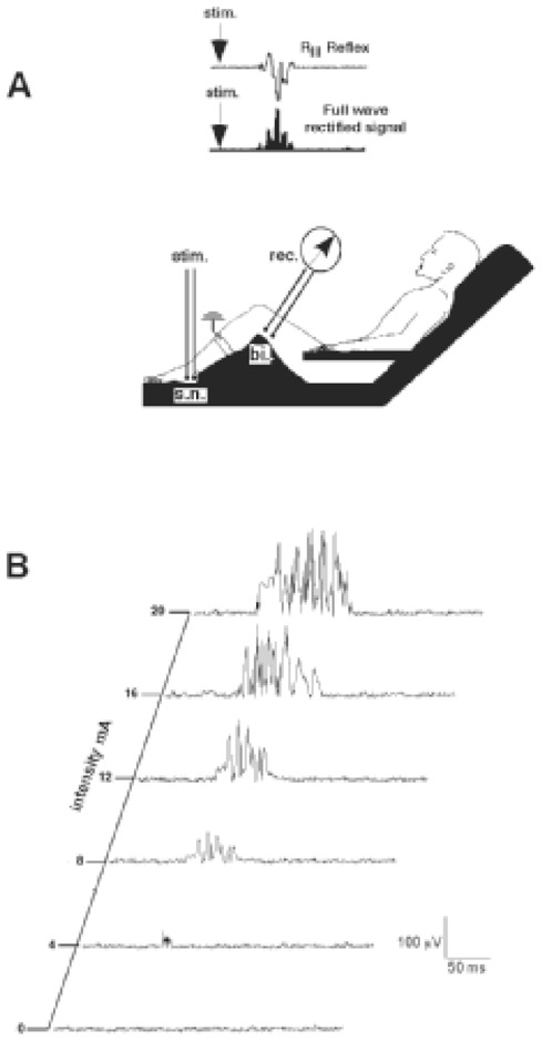
Figure 1.

A) Experimental set-up for recording the RIII reflex. The sural nerve (sn) was stimulated behind the lateral malleolus, using a pair of surface electrodes. The electrical responses were recorded (rec) from the ipsilateral biceps femoris muscle (bi) using a pair of surface electrodes. An example of an RIII reflex response, and the corresponding full-wave rectified signal, are shown in the upper part of the figure. B) individual example of RIII reflex responses with increasing intensity of stimulation of the sural nerve at 0.1 Hz.
