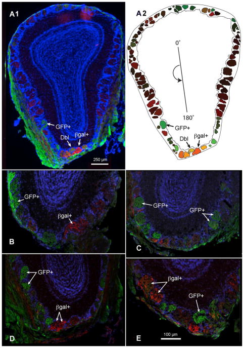
Figure 1.
Cross-sections through the olfactory bulbs of double transgenic mice expressing TrpM5-driven GFP (GFP+, green) and NT-3-driven β-galactosidase (βgal+, red). (A1) Labeled axons for both reporter genes terminate in glomeruli throughout the glomerular layer of the MOB. Arrows indicate a few strongly labeled for the two reporters: GFP+ or βgal+; Dbl − indicates a double-labeled glomerulus. (A2) Plot of each mapped glomerulus from the bulb cross-section in (A1). The origin line in the center of the plot indicates the counter-clockwise cylindrical coordinates of each glomerulus, where 0, 90, 180, and 270° indicate dorsal, lateral, ventral, and medial, respectively. The color in each glomerulus is determined by an RGB triplet in which the red and green values correspond to the mean red and green intensities of the pixels contained within the glomerular profile, and the blue value is set to zero. (B and C) GFP+ and βgal+ glomeruli are shown in the ventral MOB of a single female mouse and (D and E) a single male mouse. Immunolabeling was performed with antibodies against GFP (green) and βgal (red), reporter proteins for TrpM5 and NT-3 expression, respectively; DAPI counterstain, blue A–E. Scale bar = 100μm.

