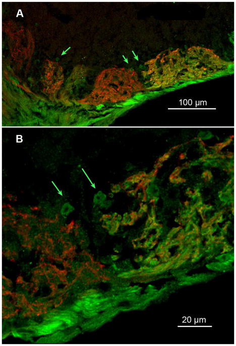
Figure 3.
(A) Confocal image (z-stack 6.0 μm deep) showing heterogeneous fields of glomerular neuropil in several glomeruli of the ventral MOB. The green arrows indicate small regions of neuropil that stain differently than the main body of the glomerulus. (B) Higher magnification of the region in panel A indicated by the paired arrows on the right side of (A) but shown in a z stack of 0.5 μm. Small round regions of TrpM5-GFP-positive neuropil are apparent at the margins of two neighboring glomeruli, one exhibiting dual label and the other being predominantly occupied by fibers exhibiting NT-3-βgal staining. A median digital filter was applied to reduce background pixel noise.

