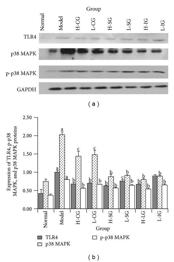
Figure 5.

Western blot analysis of proteins involved TLR4, p-p38 MAPK, and p38 MAPK in Kupffer cells (a). Expression of TLR4, p-p38 MAPK, and p38 MAPK proteins in Kupffer cells (b). Rats were fed with normal chow diet or HFD with or without CSS and SLBZSs for 16 weeks. KCs were isolated and identified from 6 rats in each group. Values represent the mean ± S.E.M. a P < 0.01 versus normal group; b P < 0.01, c P < 0.05 versus model group.
