. 2014 Mar;16(1):11–27. doi: 10.31887/DCNS.2014.16.1/rduman

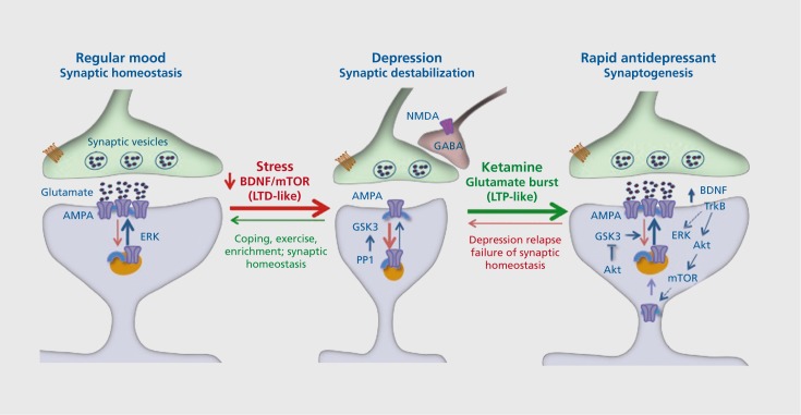
Figure 2. Stress and depression decrease, while rapid-acting antidepressants (eg, ketamine) increase, synaptic connections. Under normal, nonstress conditions, synapses of glutamate terminals are maintained and regulated by circuit activity and function, including activity-dependent release of brain derived neurotrophic factor (BDNF) and downstream signaling pathways. Stress and depression are associated with neuronal atrophy and decreased synaptic connections in the prefrontal cortex and hippocampus. This is thought to occur via decreased expression and release of BDNF as well as increased levels of adrenal glucocorticoids. This decrease has been compared with long-term depression (LTD). Rapid-acting antidepressants, notably ketamine, cause a burst of glutamate that results in an increase in synaptogenesis that has been compared with long-term potentiation (LTP). The increase in glutamate is thought to occur via blockade of N-methyl-Daspartate (NMDA) receptors located on inhibitory γ-aminobutyric acid (GABA)--ergic neurons, resulting in disinhibition of glutamate transmission. The burst of glutamate increases BDNF release and causes activation of mammalian target of rapamycin (mTOR) signaling, which then increases the synthesis of synaptic proteins required for new spine synapse formation. These new connections allow for proper circuit activity and normal control of mood and emotion. However, the new synapses are unstable and are lost after about 10 days, which coincides with depression relapse in patients. Akt, protein kinase b; AMPA, α-amino-3-hydroxy-5-methyl-4-isoxazolepropionicacid; ERK, extracellular signal-regulated kinases; GABA, γ-aminobutyric acid; GSK, glycogen synthase kinase; PP1, phosphoprotein phosphatase 1; TrkB, tropomyosin receptor kinase B.

Figure 2.