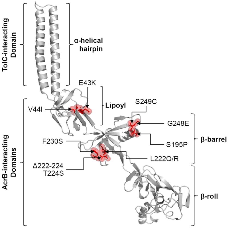
Figure 3.

Locations of AcrA suppressor alterations. A cartoon showing the model of full-length AcrA based on the partial X-ray structure of AcrA (2F1M) and the full-length MexA structure (2V4D). Locations of AcrA substitutions, which compensate for the defect of an AcrB β-hairpin mutant, are shown in red with sticks and transparent space-fill representations. AcrA residue numbering corresponds to that of the mature protein. Positions of the four AcrA domains and those that interact with TolC and AcrB are shown.
