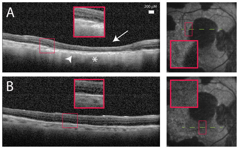
Figure 4.
A) Spectralis optical coherence tomography (OCT) image of the proband (II:3) showed outer retinal loss (arrow) and choroidal thinning (arrowhead). This area corresponded to absent blue autofluorescence as well as increased OCT signal transmission in the choroid indicative of retinal pigment epithelial loss (asterisk). B) Retinal areas with normal autofluorescence demonstrated improved retinal lamination and thicker choroid.

