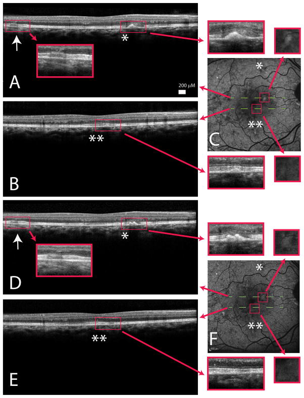
Figure 5.
A/B)Spectralis optical coherence tomography (OCT) image of a young female carrier (III:3) showed early outer retinal thickening (arrow) with focal retinal pigment epithelium hyper-reflective, drusen-like deposits (single/double asterisks) associated with the loss of the inner segment/outer segment junction. C)Autofluorescence images demonstrated macular irregularity and areas of drusen-like deposits that appeared hyper-autofluorescent. D/E)Spectralis OCT of III:3 one year later showed persistent outer retina thickening (D; arrow) and also dynamic changes with both worsening (comparing A/D asterisks) and improvement (comparing B/E double asterisks) of drusen-like material. Choroid and choriocapillaris appeared relatively normal. F)Autofluorescence obtained one year later showed increase in size of the drusen-like material correlating to an enlarged area of hyper-autofluorescence (comparing top line C/F) and resolution of the drusen-like material corresponding to a lessening of hyper-autofluorescence (comparing bottom line C/F). Red arrows showed enlarged images of highlighted areas.

