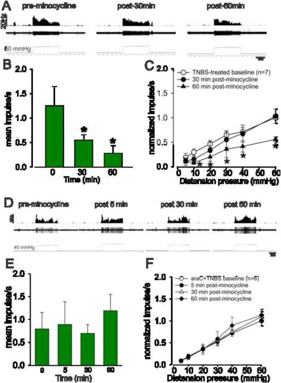
Fig.6.
Effect of minocycline on the mechanotransduction of PNA fibers from TNBS-treated rats. A: Example of the typical response of a CRD-sensitive PNA fiber from a TNBS-treated rat. In all panels, the top trace shows the response to CRD represented as a frequency histogram (1s binwidth), the middle trace is the neuron action potential and the bottom trace is the distension pressure. B: administration of minocycline (25mg/kg, i.v.) significantly decreased the spontaneous firing of these fibers. C: the drug also significantly attenuated the mechanotransduction of PNA at 60 min, but not at 30 min after injection. Values expressed as normalized mean impulse ± S.E.M of ‘n’ neurons. *P<0.05 vs TNBS-treated baseline. Effect of minocycline on the mechanotransduction of PNA fibers from araC + TNBS-treated rats. (D) Example of the typical response of a CRD-sensitive PNA fiber from araC + TNBS-treated rat. In all panels, the top trace shows the response to CRD represented as a frequency histogram (1s binwidth), the middle trace is the neuron action potential and the bottom trace is the distension pressure. (E) Administration of minocycline (25mg/kg, i.v.) did not alter the spontaneous firing of these fibers. (F) Minocycline also did not attenuate the mechanotransduction of PNA at time points investigated. . Values expressed as normalized mean impulse ± S.E.M of ‘n’ neurons.

