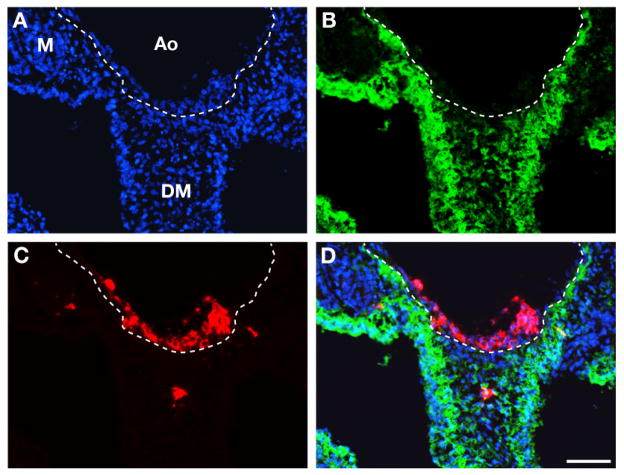
Figure 3. Tracing splanchnopleural mesoderm derivatives using CFDA-SE.
24h after CFDA-SE labeling. Same section triple stained with DAPI (A), CFDA-SE (B) and CD45 (C). The dotted line indicates the limit between the endothelium and the sub-aortic mesenchyme.
DAPI staining reveals the topography of the tissues. CFDA-SE stains the sub-aortic mesenchyme and the coelomic epithelium. CD45 stains the aortic clusters. (D) merge of DAPI, CFDA-SE and CD45 signals. The aortic HC clusters were never green in keeping with an endothelial origin of HC clusters and the complementary roles of aortic endothelium and sub-aortic mesenchyme in aortic hematopoiesis. Bar=80μm.

