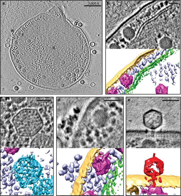
Figure 1. Zernike phase contrast cryoET enables direct recognition of cellular components of the Syn5-infected WH8109 cells.
(a) Section view of a Syn5 infected cell at late stage of infection with components labelled, including ribosomes (R), thylakoid membranes (T), carboxysomes (C), and infecting phages (I). Section and 3D annotated view of above cellular components are shown in (b) – (e). (b) Thylakoid membrane (green). (c) Carboxysome (blue). (d) Ribosome (purple). (e) An infecting Syn5 phage (red) positioned normal to the surface of the infected cell. Yellow - cell envelope; magenta - phage progeny. Panels (b) – (e), scale bars = 500Å.

