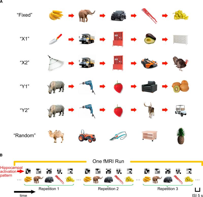
Figure 1. Object Sequences and Schematic of Sequence Retrieval.
(A) Illustration of the six types of temporal sequences. On each repetition of the “Random” sequence, the five objects were presented in a different random order, in contrast to other sequences in which the temporal order between objects was always fixed. Participants learned these sequences to criteria before proceeding to sequence retrieval session (see also Supplemental Experimental Procedures for details).
(B) Schematic diagram of sequence retrieval in one fMRI run (five fMRI runs in total). Each type of temporal sequence was presented three times within an fMRI run, with the constraint that a specific sequence was not presented consecutively and all six sequences must have been presented before the second and the third repetitions. Although brackets are shown to denote each sequence in a run, there were no explicit cues to mark divisions between sequences and the interval between objects was constant across all trials. Above each trial, a matrix is shown depicting a hypothetical hippocampal voxel activation pattern. These voxel patterns were then used to estimate similarity in hippocampal ensemble activity across different pairs of trials.

