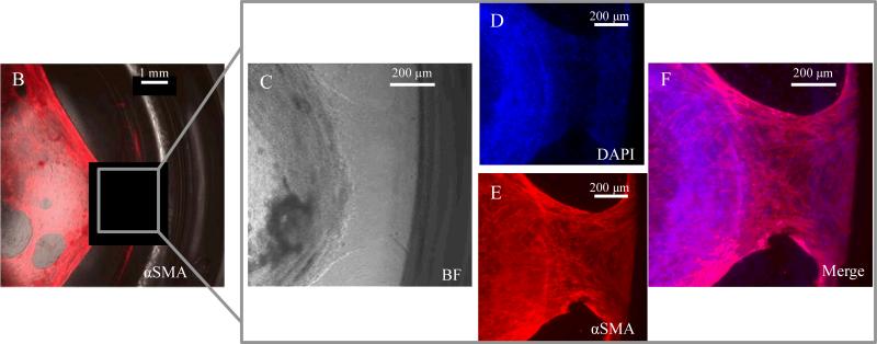
Figure 8.
A). Table of culture geometries analyzed and instances in which in vitro contraction was observed. B-F) Immunocytochemical staining of α-smooth muscle actin and nuclear DAPI staining demonstrate attachment of intestinal myofibroblast network to surrounding well edges. This observation of myofibroblast anchoring was only seen in cultures with gel diameters equal to or less than 15.5 mm. Similar to contraction, this was not observed in cultures of diameters greater than or equal to 30 mm. A minimum of 60 organoids were analyzed per condition.

