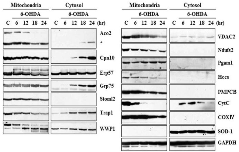
Fig. 1.
Immunoblot analysis of the identified protein expression in the mitochondrial and cytosolic fractions. MN9D cells were treated with or without 100µM 6-OHDA for the indicated time periods and subjected to cellular fractionation (mitochondria and cytosol). The fractionated lysates were separated on 8~12% SDS-PAGE and subjected to immunoblot analyses using antibodies that specifically recognize corresponding proteins. Anti-cytochrome c antibody (CytC) was used as a positive control in that release of cytochrome c into the cytosol was induced during 6-OHDA-mediated apoptosis. The following antibodies were utilized as controls; COXIV (mitochondrial marker), SOD-1 (cytosolic marker), and GAPDH (loading control).

