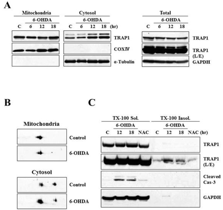
Fig. 2.
Characterization of TRAP1 release in MN9D cells following 6-OHDA treatment. (A) MN9D cells were treated with or without 100 µM 6-OHDA for the indicated time periods and subjected to cellular fractionation. Total and fractionated lysates were separated on SDS-PAGE and analyzed for TRAP1 expression using anti-TRAP1 antibody. Anti-α-tubulin antibody was used as a cytosolic marker. (B) MN9D cells were treated with 100 µM 6-OHDA for 18 hr and the fractionated samples were separated on mini 2-DE (7-cm strip) and analyzed for TRAP1 expression with the specific antibody. (C) Cellular lysates obtained from MN9D cells treated with or without 100 µM 6-OHDA in the presence or the absence of 2 mM N-acetylcysteine (NAC). Cells were then subjected to separate 1% Triton X-100-soluble from Triton X-100-insoluble fractions. Blots were probed with mouse anti-TRAP1 antibody. Anti-caspase-3 antibody was used to demonstrate drug-induced apoptotic death.

