
Figure 2. Blockade of IL-25 during RSV infection in Balb/c mice attenuates goblet cell-associated pathology and mucus production.
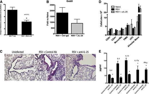
Naive Balb/c mice were infected with RSV (1×10⁵ PFU/mouse), treated with control or anti-IL-25 polyclonal rabbit anti-mouse antibody on Days 0, 2, 4, and 6, and harvested on Day 8. (A) AHR and (B) mucus-associated pulmonary gob5 expression after 8 days of infection. Cab, control antibody; Ctrl, control. (C) Histologic assessment of PAS-stained lung sections illustrates a reduction in mucus-stained airways (pink). (D) Flow cytometric assessment of individual immune cell populations in lungs of infected mice depicts no reduction in cell accumulation. Mono, Monocyte; Mϕ, macrophage; cDC, conventional dendritic cell; Macs, macrophages. (E) RSV-restimulated lung draining LN culture supernatants were assayed by Bioplex for cytokine levels. Data represent mean ± se from five mice/group. *P < 0.05 and is representative of three repeat studies.