
Figure 4. RSV-induced exacerbation of allergic airway responses is attenuated in IL-17RB−/− C57BL/6 mice.
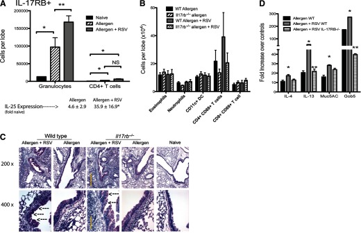
Allergic mice were infected with RSV and challenge two times with allergen (see Materials and Methods) to expose animals simultaneously to the stimuli. (A) Flow cytometry analyses indicated that the primary IL-17RB+ cell population that was increased was granulocytes. (B) Analysis of all leukocyte subsets indicated no significant change in any individual population. (C) Histological examination reflected the flow cytometry analysis in (B) indicating similar inflammatory cell accumulation in IL-17RB−/− mice; however, a reduced intensity in the PAS-postitive staining was evident in the RSV-exacerbated animals compared with WT. Arrows indicate PAS-stained goblet cells. (D) Quantitative PCR analysis of whole lung mRNA indicated that RSV-exacerbated IL-17RB−/− animals demonstrated a reduction in IL-13 and gob5 mucus gene expression. Muc5AC, Mucin 5AC. Data represent mean ± se from five mice/group.*P < 0.05; **P<0.01. This study was repeated twice with similar results.