
Figure 9.
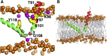
(A) Electrostatic and hydrogen-bonding interactions between caveolin-1 and surrounding lipids. (B) Membrane thinning induced by Cav182–136 insertion. Both snapshots are taken from system cav1_65_3. Cav182–136 is shown in cartoon representation; CSD domain (red). Phosphate atoms in the bulk bilayer are shown as orange spheres and phosphate atoms that are within 4.5 Å to the protein are shown in magenta. The polar and charged residues from the CSD domain (T91, K96, Y97, and R101 shown in yellow) form interactions with the lipids in the top leaflet. A water-bridged hydrogen bond between the backbone atoms of G108 (cyan spheres) and a lipid headgroup in the bottom leaflet is also shown. To see this figure in color, go online.