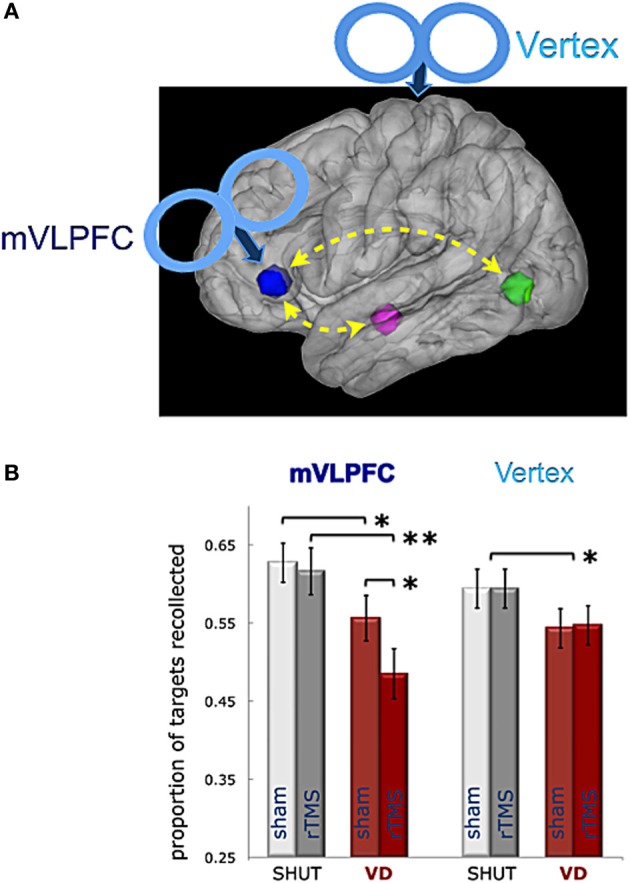
Figure 4.

Role of left VLPFC in episodic retrieval during visual distraction. A schematic illustration (A) shows the rTMS targets located on a sagittal rendering of the MNI template brain, including the mVLPFC ROI (blue) functionally connected in a recollection network with the left hippocampus (magenta) and lateral occipital cortex (green), as represented in Wais et al. (2012a). (B) The mean proportion of targets given the correct count is shown in each experiment by condition (SHUT, VD) after sham or actual rTMS treatment. Results show an interaction of actual rTMS on episodic retrieval during visual distraction after VLPFC treatment, but not after Vertex treatment. Error bars represent the standard error of the mean; ** indicates a difference between means p < 0.005; and * indicates a difference between means p < 0.05.
