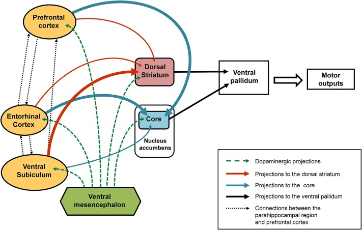
Figure 2.
Schematic representation of the connections affected following early-life (PND8) TTX inactivation. After TTX blockade of the entorhinal cortex, ventral subiculum or prefrontal cortex performed at PND8 a number of interconnected brain regions are affected (see solid arrows). Our data showed that the ventral subiculum exerts strong control over the dopaminergic responses recorded in the dorsal striatum (thick orange arrows) and only slightly affects the dopaminergic variations in the core part of the nucleus accumbens (thin blue arrow). By contrast, strong control over the dopaminergic responses recorded in the core (thick blue arrows) is exerted by the entorhinal cortex and the medial prefrontal cortex, whereas both these regions have little influence over the dopaminergic responses recorded in the dorsal striatum (thin orange arrows). The broken green arrows denote dopaminergic projections from the ventral mesencephalon. We propose that the behavioral responses observed in latent inhibition are the result of functional complementarity at the level of the ventral pallidum (dorsolateral part), of dopamine-regulated efferents from the dorsal striatum and the core part of the nucleus accumbens

