Figure 2.
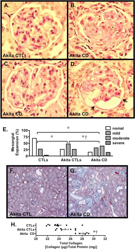
Glomerular histopathology in Akita mice. (A-D) Photomicrographs from an Akita CTL (A) and Akita Cd mice (B-D). Mild to moderate mesangial expansion was detected in glomeruli from both groups of Akita mice, which was qualitatively more severe in the Akita CD animals. Rare capsular adhesions were detected in a few mice (arrows). (E) Mesangial expansion was graded using a semi-quantitative scale as described in the Methods Section. There was a significant increase in the distribution of the semi-quantitative mesangial scores of individual glomeruli in both groups of Akita mice compared to CTLs. In Akita CD mice, there was a significant increase in the mesangial score compared to Akita CTLs. (F-G) Kidney sections stained with Masson trichrome to highlight fibrotic areas (green). A few Akita CD mice demonstrated focal areas of interstitial fibrosis. (H) Total collagen content in kidney sections was quantitated using Sirius Red/Fast Green collagen staining. There was a significant increase in collagen content in Akita CD mice compared to either Akita CTLs or CTLS. (A-D) Tissue sections were stained with Periodic acid-Schiff (PAS). (F-G) Tissue sections were stained with Masson trichrome. For the routine histological studies, 7 Akita CTLs, 9 CTLs and 5 Akita CD mice were studied. *P<0.01 vs. CTLs, †P<0.01 vs. Akita CTLs

