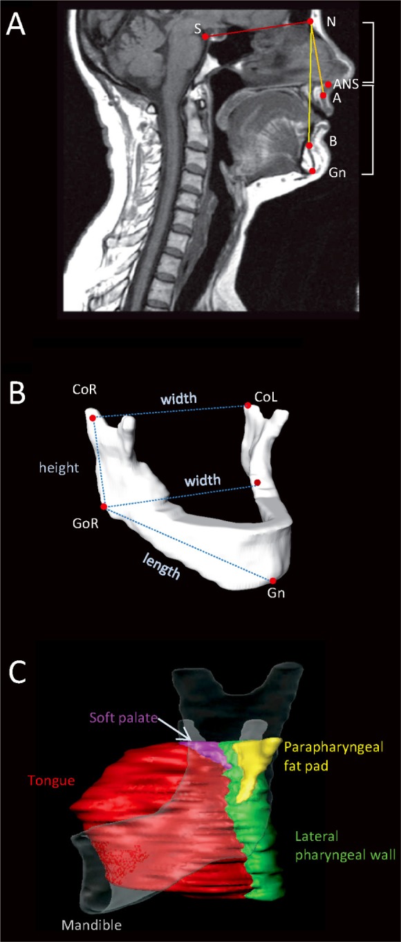
Figure 2.

Magnetic resonance imaging (MRI) analysis of craniofacial skeletal measurements and upper airway soft tissues. (A) Midsagittal MRI slice with cephalometric points: nasion (N), sella (S), anterior nasal spine (ANS), subspinale (A), supramentale (B), Gnathion (Gn). These points were used to obtain measures of sella-nasion-subspinale (SNA) and sella-nasion-supramentale (SNB) of maxillary and mandibular position and upper (N-ANS) and lower (ANS-Me) facial heights. (B) Three-dimensional reconstruction of a mandible from MRI illustrating mandibular measurements used for analysis. Right and left gonion (GoR, GoL) and condylion (CoR, CoL) and gnathion (Gn) points were identified on axial MRI slices. Three-dimensional measurements of mandibular length (Go-Gn), ramus height (Co-Go), and mandibular widths at the levels of the gonion and condyle were calculated from these landmarks and are represented by the dotted lines in the image. (C) Volumetric soft-tissue analysis. Three-dimensional reconstructions of the tongue (red), soft palate (purple), parapharyngeal fat pads (yellow), and lateral pharyngeal walls (green). The mandible is shown transparently to give perspective. Airway space volume also was calculated.
