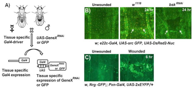
Fig. 2.

Application of the Gal4-UAS system for live imaging of wound responses and genetic screening. (a) A schematic overview of the Gal4-UAS system in Drosophila. When flies (females shown here) carrying a tissue-specific Gal4 driver are crossed to flies (males shown here) with a transgene under UAS control (for example, UAS-GeneX RNAi or UAS-GFP), progeny containing both genetic elements are produced leading to expression of the UAS-transgene in the tissues that express Gal4. (b ) Live whole mounts of the pinch-wounded larval progeny obtained after mating w; e22c-Gal4, UAS src-GFP, UAS DsRed2-Nuc/+ (e22c-reporter) flies with either w1118 (middle panel) or UAS-bskRNAi (right panel: to silence jun N-terminal kinase (JNK)) flies. The unwounded w; e22c-Gal4, UAS src-GFP, UAS DsRed2-Nuc/+ larvae are used as controls (left panel). Twenty-four hours after wounding the w1118 larvae show complete wound gap closure, but disorganized epidermal tissue (indicated by arrows) compared to the unwounded control. The larvae with epidermal JNK knockdown show open wounds indicated by the dark gap in the epidermal sheet (arrows). (c) Live larval whole mounts of the reporter line w, Nrg-GFP;; PxnGal4, UAS-2xEYFP/+ can be used to study the wound-induced inflammatory response, where a robust accumulation of hemocytes (green) labeled with YFP at the wound site can be seen 6 h after pinch wounding (right panel, compare to control)
