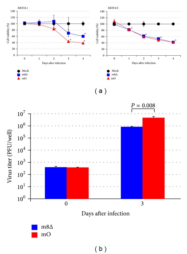
Figure 1.

Viability of FPM1 cells infected with attenuated VVs. (a) FPM1 cells were exposed to m8Δ (■), mO (▲), or PBS (●) at indicated MOI for 2 hrs. After extensive wash, the cells were cultured for indicated periods and the cell growth was assessed by using cell counting kit 8. The cell viabilities are expressed as percentages of the cell survival of mock-infected cultures. The data are presented as mean ± SD of triplicate wells. Asterisks indicate statistical significance (P < 0.05) compared to the mock-infected controls. (b) The proliferation of VVs in FPM1 cells infected with the virus at MOI 0.1 was determined by titrating the cell lysates collected at indicated days. The data are presented as mean ± SD of triplicate wells. Statistical significance was determined as P < 0.05. Similar results were obtained in two independent experiments.
