
Figure 5.
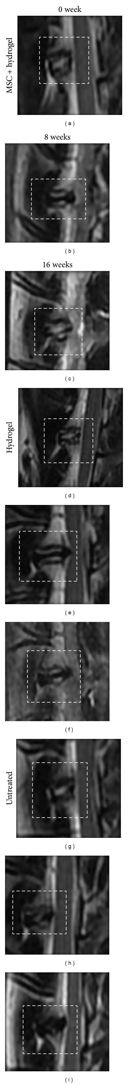
MRI images (T2 weighted sagittal views) before operation, 8 weeks, and 16 weeks after annular puncture was created. MSCs containing hydrogel group I baseline (a), 8 weeks (b) and 16 weeks (c), showing improvement in signal intensity suggesting regeneration of IVD. Hydrogel only group II baseline (d), 8 weeks (e) and 16 weeks (f), showing progression of disc degeneration. Untreated group III baseline (g), 8 weeks (h) and 16 weeks (i), showing progression of degeneration.