Figure 1.

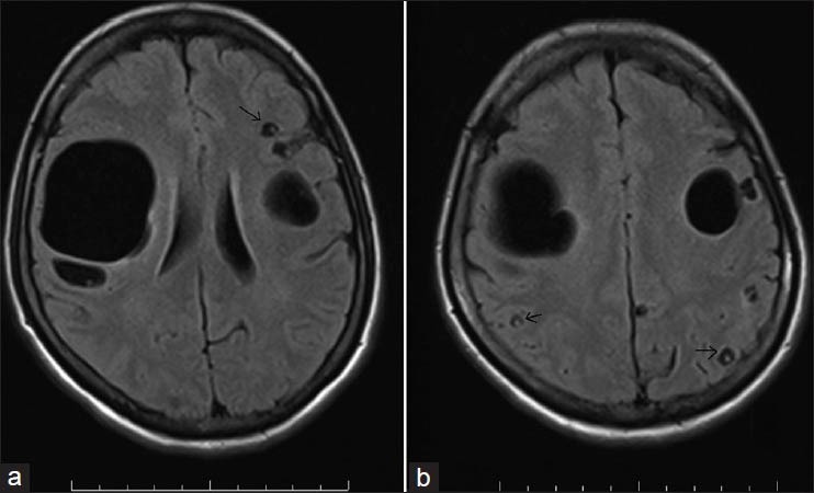
MRI brain (T1W images) showing the giant intraparenchymal cyst (a) and multiple small cysts with scolex in vesicular stage (a and b, black arrows)

MRI brain (T1W images) showing the giant intraparenchymal cyst (a) and multiple small cysts with scolex in vesicular stage (a and b, black arrows)