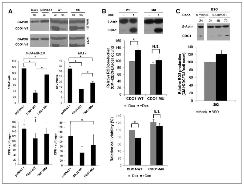
Figure 3.
Restoration of CDO1 function reduces growth and viability of cancer cells and their capacity to detoxify ROS. A, tumor cell clonogenicity was assessed on plastic and in soft-agar. Cells were transiently transfected with pcDNA3.1 (empty vector), pcDNA3.1-CDO1-WT (wild-type CDO1), or pcDNA3.1-CDO1-MU (mutant CDO1), and replated 24 hours posttransfection for selection with Geneticin/G418. After 18 days of selection, colonies were stained with Giemsa and counted. Data presented are the mean of 2 independent experiments ± SEM. Group comparisons were carried out using Student t test and trend test. *, P < 0.05. Reexpression of CDO1 was confirmed 48 and 96 hours posttransfection by Western blot analysis using α-V5 antibody, targeting the V-5-His tag of recombinant CDO1 protein, and α-GAPDH as a control. ROS production and cell viability were assayed in tetracycline-inducible CDO1-stable MDA-MB-231 cells before and after treatment with 0.5 μg/mL doxycycline (Dox; B), and 293 cells before and after the treatment with 0.5 mmol/L BSO (for depletion of glutathione; C) by fluorescence of the CM-H2DCFDA probe. Obtained values were normalized to untreated or treated empty vector controls and plotted as % relative to untreated MDA-MB-231-CDO1-WT or untreated 293 cells. Data presented are the mean of 3 independent experiments ± SEM. Group comparisons were carried out using Student t test. *, P < 0.05; n.s., not significant. Reexpression of CDO1 in MDA-MB-231 cells 48 hours after doxycycline treatment or downregulation of CDO1 expression in 293 cells 24, 48, and 72 hours after glutathione depletion with BSO was assessed by Western blot analysis using α-CDO1 antibody and α-β-actin as a control.

