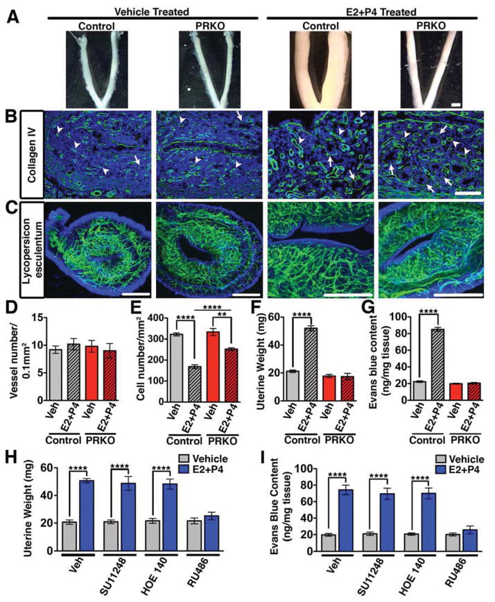
Figure 1. Reduced physiological permeability in the uterus following global PR deletion.
(A) Effect of hormones (E2+P4) on control (wild-type) and PRKO uteri. Scale, 3mm. (B) Collagen IV immunostaining (green) detects basement membrane of glands (arrows) and blood vessels (arrowheads). Scale, 100μm. (C) Uteri following intravascular perfusion with FITC conjugated Lycopersicum esculentum lectin. Scale, 1mm. (D) Vessel number/0.1mm2 in control and PRKO mice. (E) Uterine cell density/mm2 in control and PRKO mice. (F) Uterine wet weight in control and PRKO mice. (G) Uterine Evans blue content measured by the Miles assay. (H) Uterine wet weight following concurrent treatment of E2+P4 with inhibitors of VEGFR2 (SU11248), bradykinin (HOE140), and PR (RU486). (I) Quantification of Evans blue after the conditions listed in (H). In all panels, error bars = +/−. **p<0.01, ****p<0.0001. n=3–5. See also Figure S1 and Table S1.

