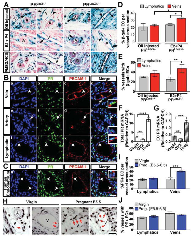
Figure 2. PR expression in the murine vasculature.
(A) β-gal positivity in transverse uterine sections from PRLacZ+/− and PRLacZ+/+ mice treated with oil, E2 and P4, or PMSG/HCG. Endothelial cells (arrowheads); smooth muscle cells (arrows); veins, V; arteries, A. Nuclear Fast Red was used as a counterstain. (B,C) Immunofluorescence of murine (B) and human (C) uterine sections stained for PECAM-1 (red) and PR (green). Nuclei were visualized using DAPI (blue). Endothelial cells (arrowheads); smooth muscle cells (arrows). Insets are higher mag images of PR positive endothelial cells. (D) Percentage of β-gal+ endothelial cells per vessel cross-section from PRLacZ+/− mice. n=3. (E) Percentage of vessels in the uterus that contain at least one β-gal+ endothelial cell per cross-section. n=3. (F) Total PR mRNA levels in the uterus from virgin, ovarectomized (OVX), and pregnant (E5.5) mice. (G) PR mRNA levels from isolated uterine endothelial cells from the same mice listed in (F). (H) PR protein expression in the endothelium of virgin and pregnant (E5.5) mice. PR+ endothelial cells (arrows); veins, V; arteries, A. (I) Percentage of PR+ endothelial cells per vessel cross-section from virgin and pregnant uteri (E5.5). n=3. (J) Percentage of vessels from virgin and pregnant uteri that contain at least one PR+ endothelial cell per cross-section. n=3. In all panels, error bars=+/−SEM. Scale=25 μm. *p<0.05, **p<0.01, ***p<0.001 ****p<0.0001. See also Figure S2.

