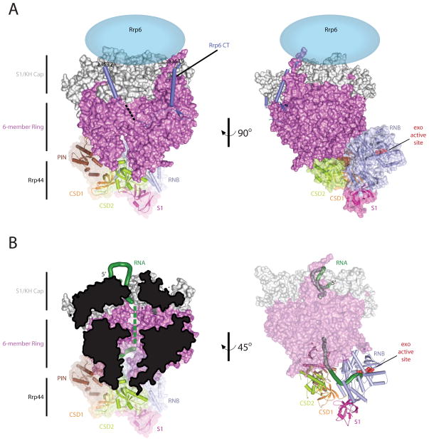
Figure 4. The 10 component exosome with CT of Rrp6.
A) Overall architecture of the Exo1144/6CTD. Orthogonal view of the budding yeast 10-subunit exosome in complex with the carboxy terminus of Rrp6 (PDB ID = 4IFD). The S1/KH cap is shown in light gray, the six-membered ring is shown in dark pink, the Rrp6 CT is shown as a light blue cartoon, the location of the EXO and HRDC are currently unknown but they are predicted to be above the cap (light blue ellipse), and each of the Rrp44 domains are colored as discussed in Fig 3. B) RNA ingress into Rrp44 exo active site. The RNA ingress via the central channel is highlighted in two different views: Left, the central channel is viewed by cutting the core in half, and right, the RNA ingress is viewed by making the core transparent.

