
Figure 3.
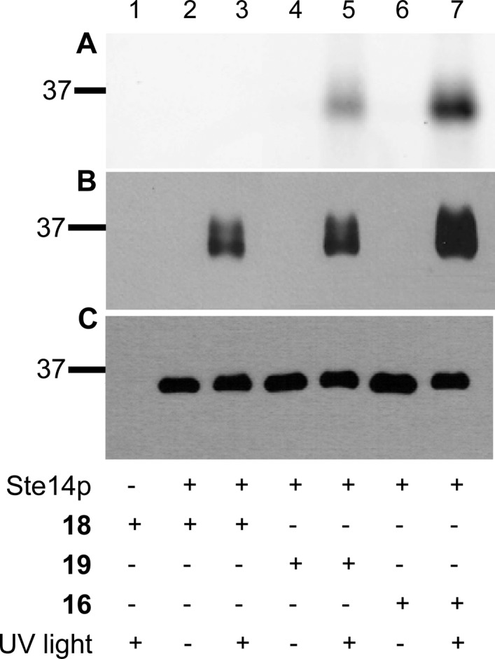
Analysis of cross-linking reactions containing purified His-Ste14p and different photoactive probes. (A) Fluorescent imaging, (B) immunoblot analysis with NeutrAvidin HRP, and (C) immunoblot analysis with α-Ste14 . Experiments were performed with 16, 18, and 19. For this experiment, purified His-Ste14p (0.25 μg) was incubated with the probes indicated (50 μM) and irradiated on ice for 30 min followed by fractionation via SDS-PAGE. The resulting gel was visualized using (A) a fluorescence scanner or transferred to a nitrocellulose membrane and visualized using (B) NeutrAvidin HRP or (C) an α-Ste14 antibody. The data shown is from one of three replicates of this experiment.