
Figure 4.
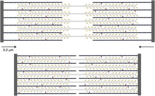
Schematic description of a cardiac sarcomere drawn to scale. (Top) A sarcomere is depicted at its resting length just beginning its contraction. The sarcomere is 2-μm long, the myosin bipolar thick filaments are 1.6-μm long, and the actin filaments are 0.8-μm long. The total number of myosin molecules that an individual actin filament sees is ∼50; only 32 are shown here because the myosin molecules from the third thick filament that is out of the plane of focus here are not shown. Note the myosin molecules are depicted as single-headed for simplicity. A duty ratio of 0.1 is depicted. Thus, ∼10% of the myosin molecules are in a force-producing state (red). (Bottom) The sarcomere has shortened by ∼20%. To see this figure in color, go online.