
Figure 6.
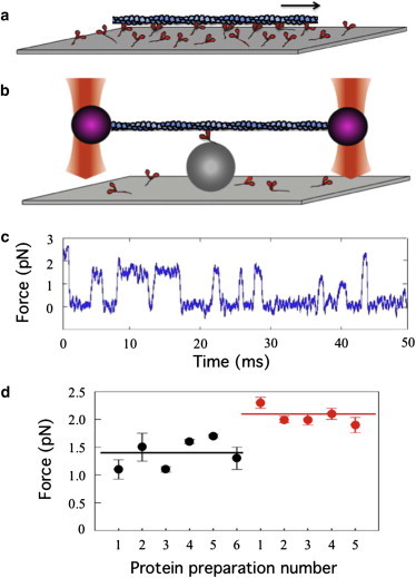
In vitro motility taken to the single-molecule level. (a) Myosin-coated surfaces drive the movement of fluorescently-labeled actin filaments at velocities comparable to those of muscle contraction (85). (b) The dual-beam laser-trap assay for measuring nanometer steps and piconewton forces of a single myosin molecule (55). (c) Force transients measured by clamping the position of the actin-bound polystyrene bead on the left (purple, in panel b) as the myosin is trying to move the actin to the right. (d) Mean intrinsic forces from multiple preparations of wild-type human β-cardiac myosin S1 (black circles) and human β-cardiac myosin S1 carrying the HCM-causing mutation R453C (red circles) (49). To see this figure in color, go online.