
Figure 4.
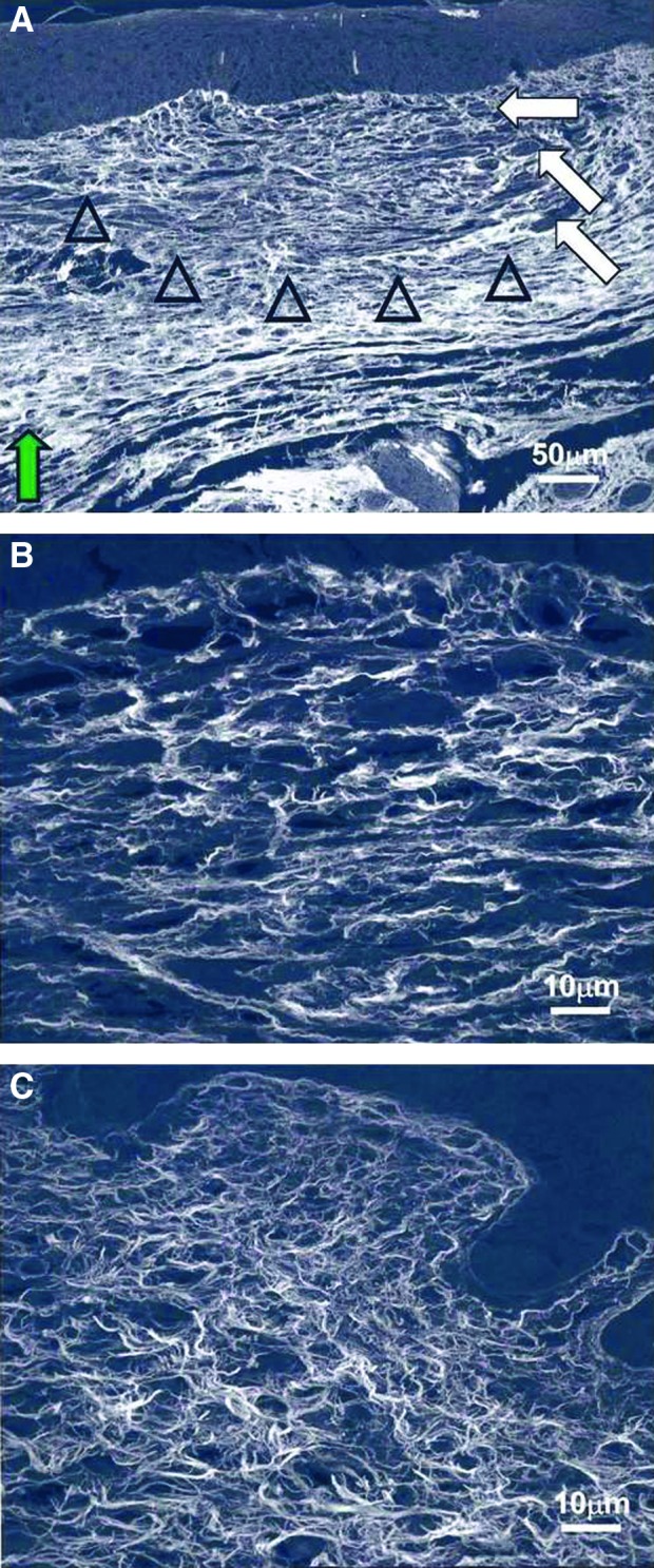
Confocal microscopy of scarring E18.5 fetal wounds. Collagen fibers are stained with Sirius red and appear white. (A) Healed wound skin harvested at 72 h (200×). The dermal collagen pattern (open arrowheads) is different from the surrounding nonwounded dermis (green arrow). The fibers are less densely compacted, and no epidermal appendages are present. Neovascularization is shown with the white arrows. (B) Healed wound at 72 h at a higher magnification (1,000×). The collagen fibers are thicker but with greater interfiber spaces compared with nonwounded dermis. (C) Nonwounded skin at 21.5 days of gestational age (1,000×). When compared with wound collagen fibers (B), nonwounded dermal collagen fibers are thinner, with less interfiber space. Reprinted with permission from Beanes et al.3 To see this illustration in color, the reader is referred to the web version of this article at www.liebertpub.com/wound