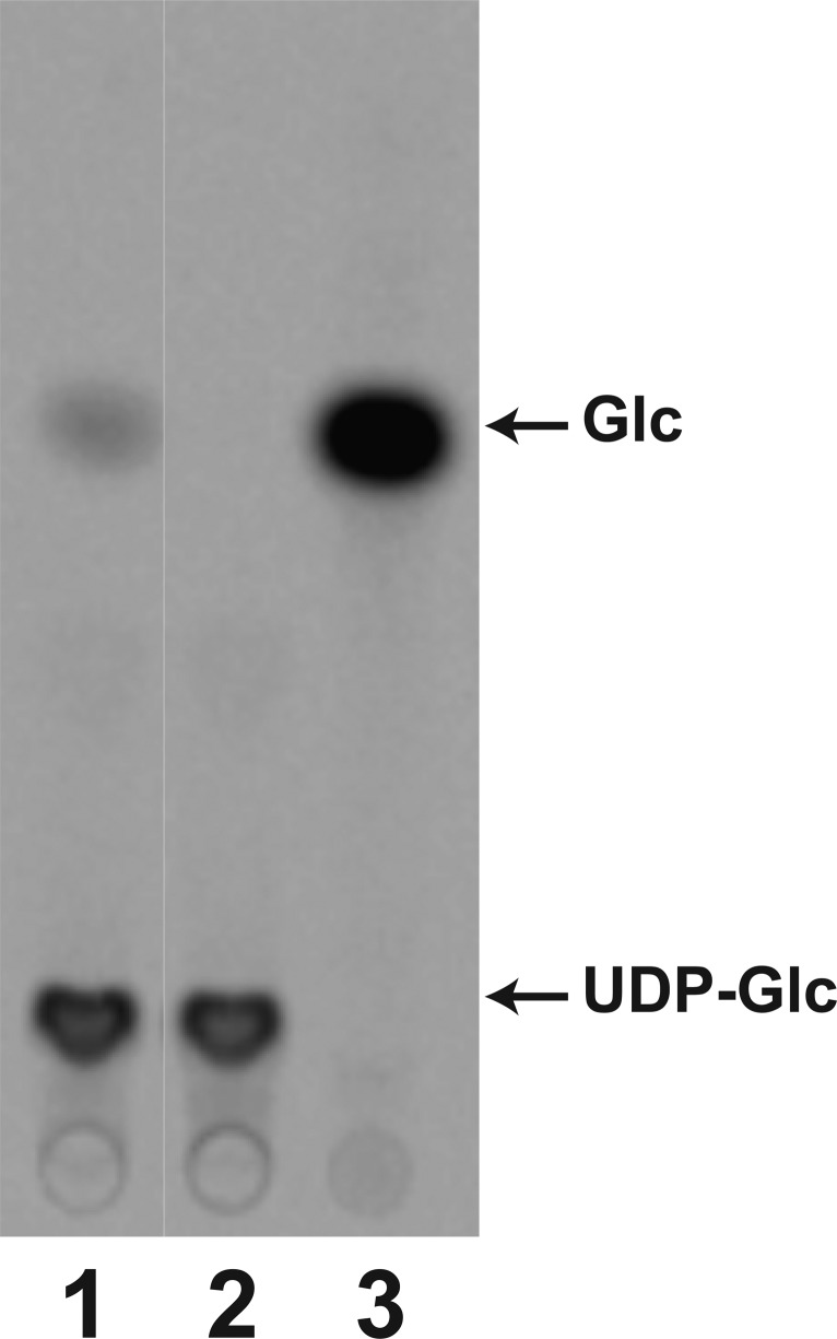
Figure 5.
WaaG hydrolyzes UDP-Glc in the absence of a lipid acceptor. UDP-[U-14C]Glc was incubated in the presence (lane 1) or absence (lane 2) of WaaG (0.25 mg/mL) and then displayed using TLC as described in Materials and Methods. [U-14C]Glc was spotted as a control in lane 3 to indicate the migration of free glucose.

