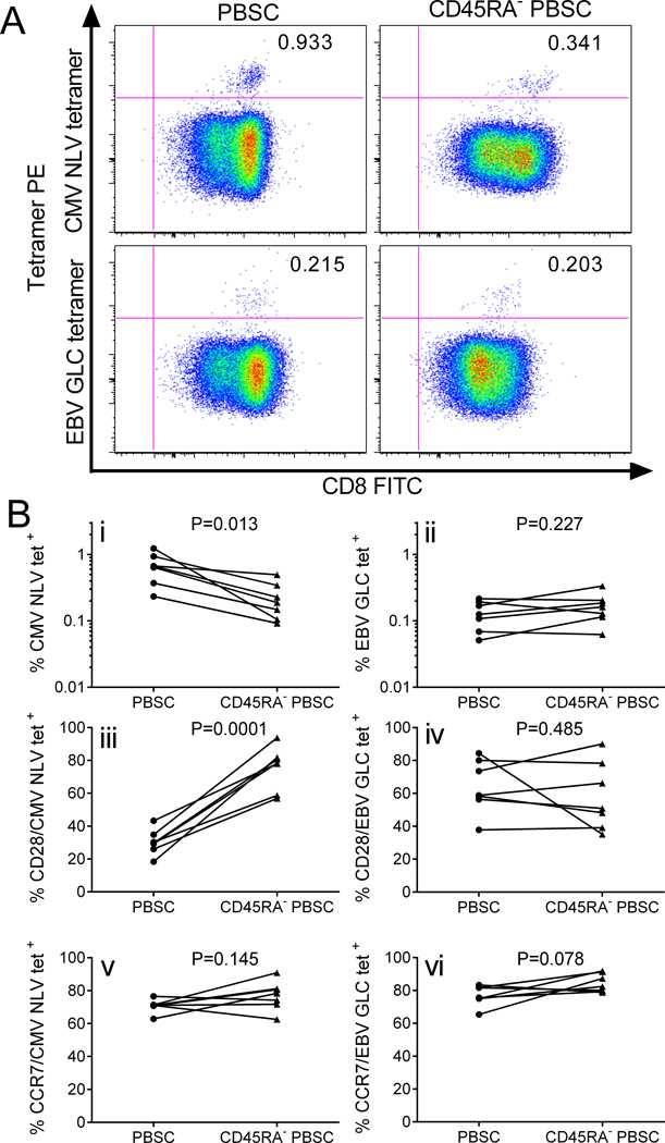
Figure 5. Virus-specific T cells in G-PBSC before and after CD45RA-depletion.
A) Flow cytometry plots showing MHC tetramer staining for viral epitopes (CMV pp65 NLVPMVATV and EBV BMLF1 GLCTLVAML) of CD8+ T cells enriched from G-PBSC and CD45RA-depleted PBSC from a representative HLA-A2+ CMV+ EBV+ donor. B) Tetramer evaluation of G-PBSC and CD45RA-depleted PBSC from 7 HLA-A2+ CMV+ EBV+ donors (i) CMV NLV specific T cells and (ii) EBV GLC specific T cells as a proportion of CD8+ T cells; CD28 expression on (iii) CMV NLV CD8+ and (iv) EBV GLC CD8+ T cells; CCR7 expression on (v) CMV NLV CD8+ and (vi) EBV GLC CD8+ T cells. Analyzed by Student’s paired t test.

