
Figure 1.
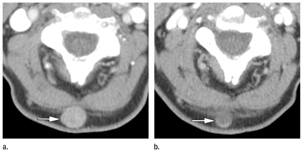
Axial CT images in a 64-year-old man with metastatic melanoma who was treated with bevacizumab antiangiogenic therapy. Patient had normal baseline serum LDH (144 IU/L) and metastases involving subcutaneous tissues, lung, pleura, mediastinal and hilar lymph nodes, adrenals, retroperitoneal fat, and bowel. (a) Pretherapy contrast-enhanced CT image shows 1.7-cm hypervascular subcutaneous metastasis (mean attenuation, 93 HU) in the posterior aspect of the neck (arrow). (b) On initial posttherapy contrast-enhanced CT image, metastasis had decreased in size (1.2 cm) and decreased in mean attenuation (30 HU). Total target lesion burden decreased by 43%. Per MASS criteria, patient had favorable response. Target lesion met criteria for both marked decreased attenuation (≥ 40 HU decrease) and marked central necrosis (> 50% of mass changing to near fluid attenuation after therapy). Patient had good clinical outcome, with progression-free survival of 22 months and overall survival of 26 months.