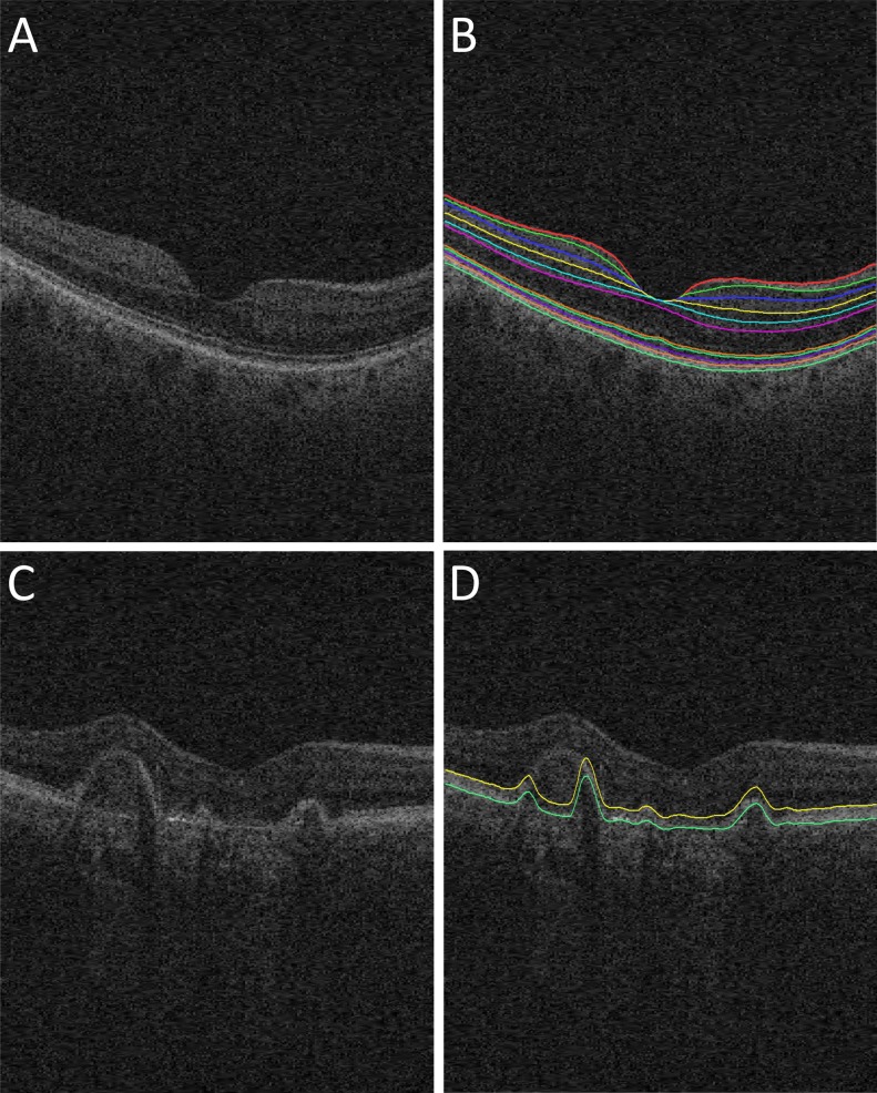
Figure 2.
Retinal surfaces segmentation. (A) Original B-scan of 3D SD-OCT image from a normal subject. (B) Eleven retinal surfaces segmentation for the normal subject.11 (C) Original B-scan of SD-OCT image from an exudative AMD patient. (D) Initial segmentation of ORSR surfaces of the exudative AMD patient, showing incorrect segmentation (yellow surface: myoid IS–ellipsoid IS; green surface: Bruch's membrane).

