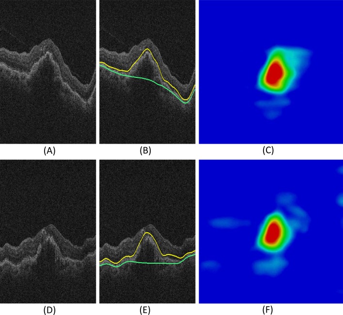
Figure 6.
Reproducibility of ORSR layer segmentation. (A–C) First visit image data of an example subject. (A) Original slice. (B) ORSR segmentation (yellow surface: myoid IS–ellipsoid IS; green surface: Bruch's membrane). (C) Thickness map of the first visit image data. (D–F) Second visit image data of the same example subject. (D) Original slice. (E) ORSR segmentation (yellow surface: myoid IS–ellipsoid IS; green surface: Bruch's membrane). (F) Thickness map of the second visit image data.

