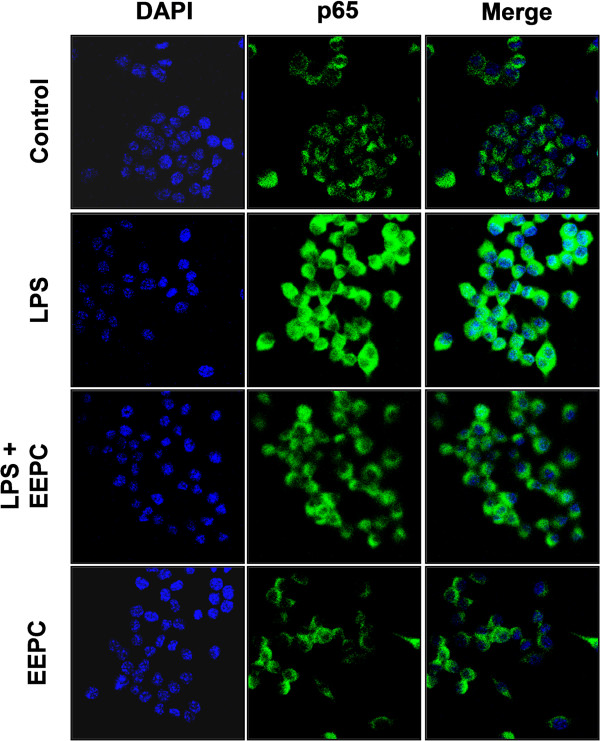
Figure 6.
Effects of EEPC on LPS-induced nuclear translocation of NF-κB p65 in RAW 264.7 macrophages. Cells were pre-treated with 8 μg/ml EEPC for 1 h, LPS (0.5 μg/mL) was then added, and cells were incubated for 30 min. Localization of NF-κB p65 was visualized with fluorescence microscopy after immunofluorescence staining with NF-κB p65 antibody (green). Cells were stained with DAPI for visualization of the nuclei (blue). A representative sample of three independent experiments is shown.

