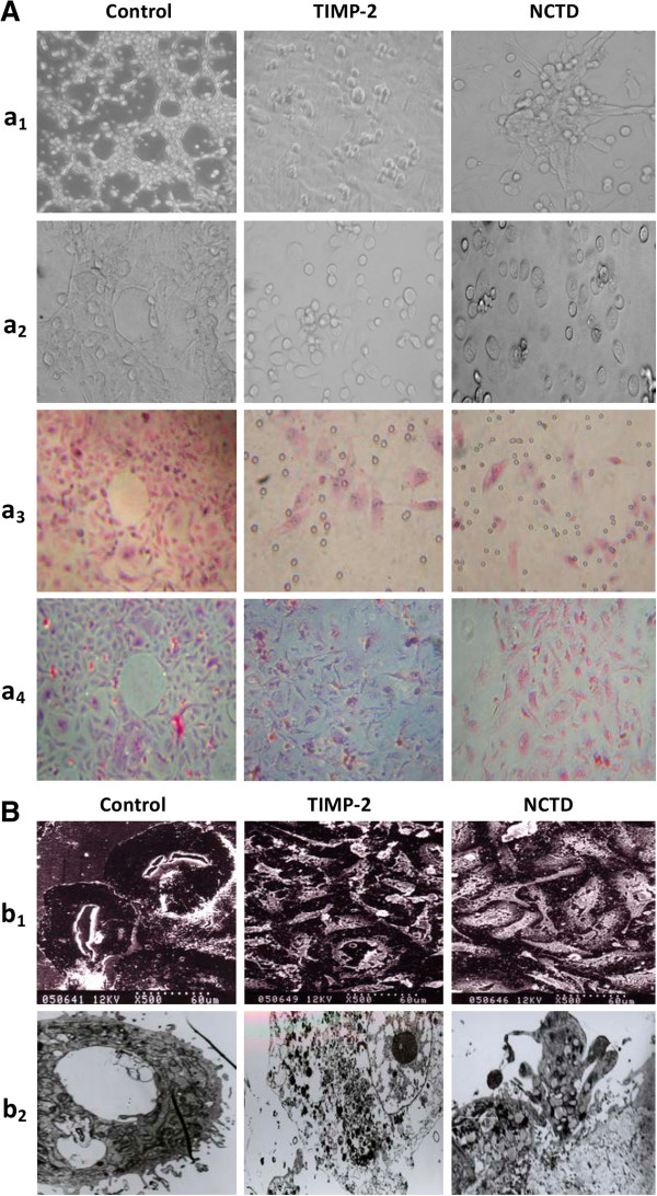
Figure 4.
Phase contrast microscopy and electron microscopy on 3-D cultures of GBC-SD cells in vitro. (A) Phase contrast microscopy of GBC-SD cells 3-D cultured on Matrigel and rat-tail collagen I matrix (original magnification, ×200) in vitro. GBC-SD cells formed patterned, vasculogenic-like networks when cultured on Matrigel (a1) and rat-tail collagenImatrix (a2-3, H&E staining; a4, PAS staining without hematoxylin counterstain) for 14 days; furthermore, PAS positive, cherry-red materials found in granules and patches in the cytoplasm of GBC-SD cells appeared around the signal cell or cell clusters. But in the process of network formation, using TIMP-2 or NCTD for 2 days, GBC-SD cells lost the capacity of the above vasculogenic-like network formation, with visible cell aggregation, float, nuclear fragmentation, apoptosis and necrosis. (B) Vasculogenic-like network microstructures in 3-D cultures of GBC-SD cells under electron microscopies (b1, SEM × 500; b2, TEM × 1200). SEM or TEM clearly visualized channelized or hollowed vasculogenic-like networks formed GBC-SD cells (red arrowhead), with clear microvilli surrounding cluster of tumor cells; also, TEM showed some microvilli on the outside of network, clear cellular organelle structures, and cell connection with an increased electron density in density (yellow arrowhead). After using TIMP-2 or NCTD for 2 days, GBC-SD cells couldn’t grow along with collagen framework, raised and deformed, lost the capacity of the above network formation (blue arrowhead), with visible decreased microvilli, destroyed cellular organelles, Vacuolar degeneration (green arrowhead), nuclear fragmentation, and typical apoptotic bodies (brown arrowhead).

