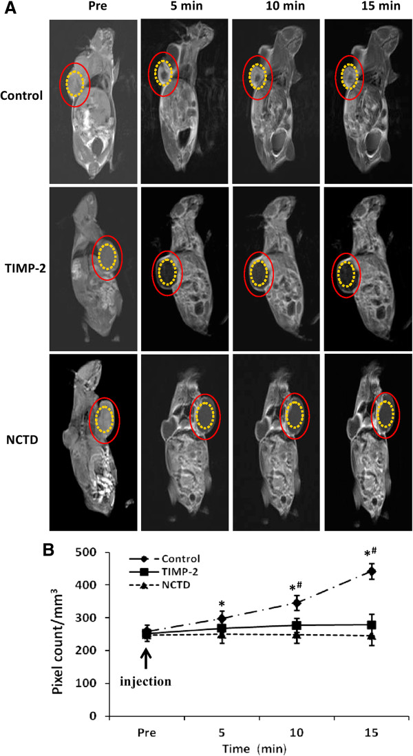
Figure 6.
Dynamic micro-MRA and hemodynamic of GBC-CD xenografts in vivo. (A) The images were acquired before the injection (pre), 5, 10, and 15 min after injection of the contrast agents (HAS-Gd-DTPA). The tumor center area (yellow circle) in control group exhibited a signal that gradually increased multiple spots in intensity (which is consistent with the intensity observed in tumor marginal area between the (red circle and the yellow circle). However, the center region (yellow circle) of the xenografts in NCTD or TIMP-2 group exhibited a decreased signal or a lack of signal change in intensity. (B) Hemodynamic changes of the xenografts’ VM of each group. All data are expressed as means ± SD. *P < 0.001, vs. Pre injection in control group; #P = 0.0000, vs. NCTD or TIMP-2 group. But, no difference on signal intensity (pixel count/mm3) was observed between NCTD group and TIMP-2 group.

