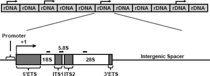
Figure 1. The qPCR-based assay to determine the genomic content of rDNA.
The rDNA copies are organized as long tandem repeats located on five acrocentric chromosomes. Each copy consists of the rRNA gene and the intergenic spacer (IGS). Each rRNA gene includes a Pol1-dependent promoter and exons that correspond to 18S-, 5.8S- and 28S rRNAs. They are separated by introns (5’ETS, ITS1, ITS2 and 3’ETS). The positions of the analyzed rDNA amplicons are indicated by the thick black lines. The schematics are not drawn in scale.

