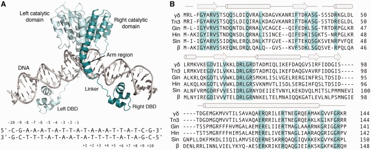
Figure 1.
Overview of the small serine recombinases. (A) (Top) Crystal structure of the γδ resolvase dimer bound to target DNA (PDB ID: 1GDT) (20). ‘Left’ and ‘right’ recombinase monomers are colored light and dark teal, respectively. DBD indicates native DNA-binding domain. Linker and arm region are labeled for the ‘right’ recombinase monomer only. (Bottom) Core sequence recognized by the γδ resolvase catalytic domain. Base positions are indicated. (B) Sequence alignment of six of the most comprehensively characterized serine recombinase catalytic domains. Conserved residues are highlighted light teal. The α-helical and β-sheet secondary structural elements are denoted above the alignment as cylinders and arrows, respectively.

