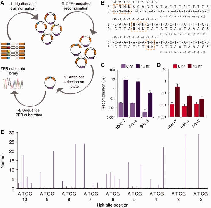
Figure 3.
Specificity of the β-N95D catalytic domain. (A) Schematic representation illustrating the genetic screen used to profile recombinase specificity. Recombinase substrate library shown in various colors; ZFR gene is in purple, β-lactamase gene is in orange and GFPuv gene is in white. (B) Randomization strategy used for specificity profiling. Randomized bases are boxed. Note that only ‘left’ half-site of the upstream ZFR target site contained base substitutions. (C and D) Recombination by (C) β-N95D and (D) Sin-Q87R/Q115R for each 20B and 20S core site library, respectively, at 6 and 16 h. (E) Number of selected base sequences (out of 30) at each position within the 20B half-site. Thirty clones were sequenced from each 6-h library output. Recombination was determined by split gene reassembly. Error bars indicate standard deviation (n = 3).

